<<<
Compare
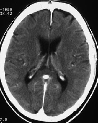
the pathological image-left and the physiological image-right
(blinded)
<<
F:
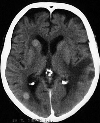
At
brain windows (CCT after contrast) are two, rounded, enhancing soft-tissue masses
near the rigth caudate nucleus and the right occipital lobe. 2) Deepening of
the sulci and narrowing of the gyri
H:
Adult
man, 72-years-old, with known bronchial carcinoma - now staging.
INFO/WWW-LINKS:
1) More than 30% of intracranial neoplastic diseases are caused by metastases/secondaries
(breast, lung, melanoma). Primaries include: astocytomas, glioblastoma multiforme,
oligodendrogliomas, ependymomas, meningiomas. 2) Cerebral degeneration: Cerebral
atrophy can be focal, especially temporoparietal in M. Alzheimer and can be
caused by multiple sclerosis and cerebrovascular
disease (multiple small cerebral infarcts).
IN
THIS PART OF THE PAGE YOU FIND SOME TEXT FIELDS WHICH CAN BE OPENED EIGTHER
STEP BY STEP (CLICK ON "HISTORY", "HELP", "FINDINGS",
"DIAGNOSIS" OR "INFO/WWW-LINKS") OR AT ONCE WITH A CLICK
ON "ALL ON" - VICE VERSA CLICK ON "ALL OFF".
If you need a physiological
image to compare click here