<<<
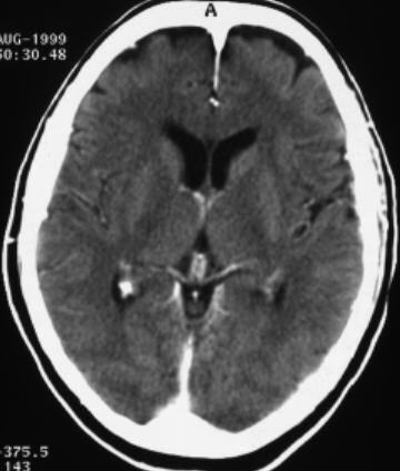
Compare
the pathological image-left and the physiological image-right
(blinded)
<<
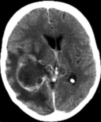
F:
1)
ring-enhancing soft-tissue mass with 2) surrounding large hypodense area and
3) central necrotic area (low density area) in the right hemisphere with 4)
ventricle compression 5) midline shift to the left side and 6) flattened sulci.
H: