<<<
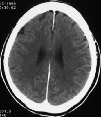
Compare
the pathological image-left and the physiological image-right
<<
Please
search for that left-sided structure or region, which is different from the
right image
IN
THIS PART OF THE PROGRAM IT IS NOT THAT MUCH IMPORTANT TO FIND THE RIGHT DIAGNOSIS.
IT IS MORE IMPORTANT TO SEE THE DIFFERENCE BETWEEN THE IMAGE ON THE LEFT AND
THE IMAGE ON THE RIGHT. THUS THE USER MAY GET A CLUE OF TYPICAL CT-FEATURES
LIKE FILLING-DEFECTS, CONTRAST ENHANCEMENT AND DENSITY.
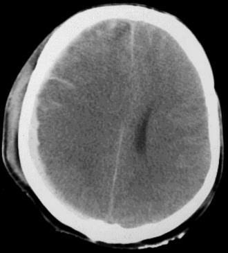
DIAGNOSIS:
1) Acute subdural haematoma
2) acute frontoparietal subarachnoid haematoma
(blood
between the gyri) 3) compression
of the right ventricle 4) midline shift
to the left side
5) Flattened sulci as evidence for brain oedema
6) Subgaleal haematoma